
Glaucoma care in New Albany.
Glaucoma damages the optic nerve gradually and silently, usually starting with your side vision. According to the National Eye Institute, it is a leading cause of blindness in the United States. With early testing and treatment, its progression can usually be slowed, which is why regular eye exams matter.
If you'd rather just pick a time, you can schedule now or call us at (614) 933-0575.
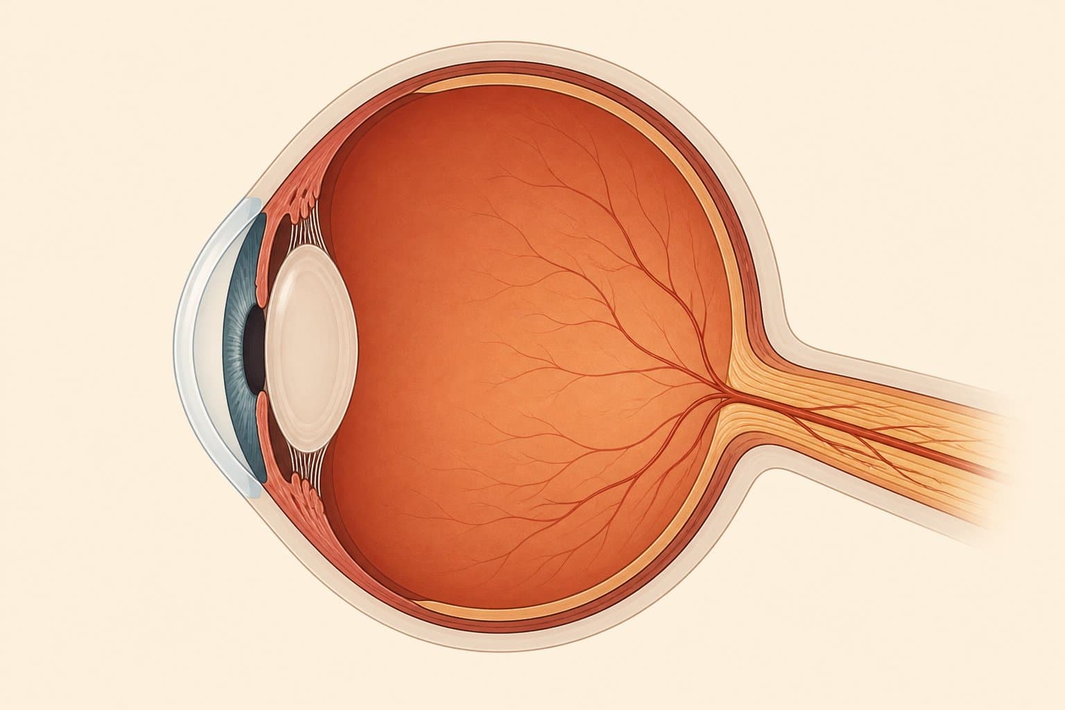
What glaucoma does to the optic nerve.
Glaucoma is a group of eye diseases that damage the optic nerve, the bundle of more than a million nerve fibers that carries everything your eye sees to your brain. In most cases the damage is tied to the pressure of the fluid inside the eye. The eye constantly makes and drains this fluid, and when the drain works poorly, pressure builds and slowly wears on the nerve. Some patients develop glaucoma at normal pressures, which is one of the reasons a thorough workup looks at the nerve itself, and at how it is functioning, over time.
The most common form, open-angle glaucoma, is painless and develops over years. It usually takes side vision first, so gradually that the brain fills in the missing areas, and central vision often stays sharp until the disease is advanced. The CDC estimates that about half of the people who have glaucoma don't know they have it. Early on there is usually nothing to feel and nothing to notice, which is why glaucoma is found at eye exams rather than from symptoms.
Vision lost to glaucoma does not come back. Treatment works by lowering eye pressure to protect the nerve fibers you still have, and with steady treatment, progression can usually be slowed. The earlier glaucoma is found, the more vision there is to protect.
Anyone can develop glaucoma, and the risk rises with age. Family history matters: having a parent or sibling with glaucoma raises your risk considerably. The American Optometric Association also lists higher eye pressure, thin corneas, diabetes, high nearsightedness, past eye injuries, and long-term steroid use among the risk factors. Risk also runs higher for Black patients beginning around age 40, and for Hispanic patients over 60. If any of these describe you, regular exams are the reliable way to stay ahead of it.
Sudden eye pain with a red eye, blurred vision, halos around lights, or nausea can mean acute angle-closure glaucoma. It is an emergency. Call us at (614) 933-0575 right away, or go to urgent care.

A complete glaucoma workup in our New Albany office.
No single test gives the full picture. We check eye pressure, the optic nerve, side vision, and the eye's drainage angle. We run all of these tests in our office. Each result is dated so your doctor can compare it with future visits. Your doctor also looks at the optic nerve directly with an exam microscope (the slit lamp) and reviews every result with your history.
iCare eye-pressure checks
Eye pressure is the one glaucoma risk factor treatment can change. We measure it at every visit. Our iCare tonometer takes the reading with a feather-light tap, with no air puff and no numbing drops. Most patients barely notice it.
OCT of the optic nerve
Optical coherence tomography (OCT) is a quick scan of the optic nerve. It measures the layer of nerve fibers that carries visual signals. Over time, these scans may show thinning before you notice any change in your vision. Nothing touches your eye.
Visual field testing
A visual field test maps your side vision point by point and finds blind spots the brain has been quietly filling in. Repeating the test over time helps show whether the disease is stable.
Optic nerve photography
Wide-field retinal photos give your doctor a detailed, dated picture of the optic nerve itself. Comparing the photos over time can reveal small changes in the nerve's shape or color.
Corneal thickness (pachymetry)
A quick measurement of the thickness of your cornea. Thin corneas raise glaucoma risk and change how pressure readings are read. This measurement helps your doctor interpret your pressure correctly.
Drainage-angle assessment (gonioscopy)
Using the exam microscope and a special lens, your doctor can see the angle where fluid drains out of the eye. Whether the angle is open or narrow helps identify the type of glaucoma and guide treatment.
Treatment and monitoring, visit after visit.
For most patients, glaucoma treatment means daily prescription eye drops that lower eye pressure. Your doctor checks how the drops affect your pressure and adjusts them toward a pressure goal set for your eye. That goal may change as new OCT and visual field results come in. We treat and monitor many glaucoma patients right here in the office.
Glaucoma care has a rhythm. Between full exams, shorter visits check your pressure. Your doctor repeats OCT and visual field tests based on how stable your results are. Each new result is compared against your baseline. A single test can vary. Your doctor checks for change by reading pressure, OCT, visual fields, and the exam together.
Some glaucoma needs more than drops. If glaucoma advances despite treatment, laser treatment or surgery may be the next step. We refer you to an ophthalmologist (an eye surgeon) and coordinate your care. Your routine eyecare stays here, with the doctors who know your history.
Glaucoma visits are medical eyecare. They are usually billed to medical insurance, not a vision plan. We accept many medical plans, including Anthem, Aetna, Cigna, Medicare, UnitedHealthcare, and Medical Mutual. We verify your coverage before your visit.
Medicare may cover medically necessary glaucoma exams and follow-up visits. If you're on Medicare, our Medicare eye exams page walks through how that coverage works. Call us at (614) 933-0575 with your insurance card and we'll confirm the details together.
Glaucoma questions we hear in the exam room.
Q.01 Can vision lost to glaucoma come back? +
No. Damage to the optic nerve is permanent, which is why finding glaucoma early matters so much. Treatment lowers eye pressure. That can usually slow further damage and help protect the vision you still have.
Q.02 Does glaucoma have early warning signs? +
Usually none. The most common form is painless and slow. Side vision may fade so gradually that the brain fills in the gaps. For most patients, a comprehensive eye exam is how glaucoma is found.
A rare exception is acute angle-closure glaucoma. It starts suddenly, with intense eye pain, a red eye, blurred vision, halos around lights, and sometimes nausea. That is an emergency. Call us right away, and if it is after hours, seek emergency care.
Q.03 Does the eye pressure check hurt? +
No. We measure pressure with the iCare tonometer, which touches the eye for a fraction of a second with a tiny, feather-light probe. There is no air puff and no numbing drop, and most patients barely feel it.
Q.04 What does it mean to be a "glaucoma suspect"? +
It means your doctor found something worth watching. That might be higher pressure, an unusual optic nerve shape, or a strong family history. Many glaucoma suspects never develop glaucoma. OCT, visual fields, and pressure checks help us tell whether glaucoma is developing. If treatment is ever needed, it can start promptly.
Q.05 Can COVE manage my glaucoma, or will I need a specialist? +
Our doctors diagnose and treat glaucoma here in New Albany. We prescribe drops and track the optic nerve over time. If you need laser treatment or surgery, we refer you to an ophthalmologist and coordinate your care.
Q.06 How often will I be seen? +
It depends on how stable your glaucoma is. Most patients in treatment come in several times a year for pressure checks. Your doctor sets the schedule for visual field and OCT tests. We set the interval with you and adjust it as your results come in.
Schedule an exam that checks your optic nerve.
Every comprehensive eye exam at COVE includes glaucoma screening, which typically includes an iCare pressure check, retinal photos, and a screening OCT. Schedule online or call (614) 933-0575. If it has been more than a year since your last exam, this is a good reason to come in.
Related: diabetic eye exams · macular degeneration · routine eye exams · COVE Plus flat pricing for uninsured patients